1. Introduction
Urine is a complex solution generated in the kidney of humans and animals through the metabolism of endogenic wastes, drinks, drugs and foods [1]. The process through which urine is generated in the kidney is known as urinalysis. The composition and properties of urine vary with the source, organism's feeding habits, body size, amount of water consumed, environmental factors [2,3] and the health-condition of the organism that released the urine [4]. It is composed of diverse inorganic compounds such as potassium, phosphorus, nitrogen, and sodium, as well as organic compounds including creatinine, creatine, and uric acid, as major components [5]. The different components in urine are basically influenced by the composition of essential elements in the urine [6].
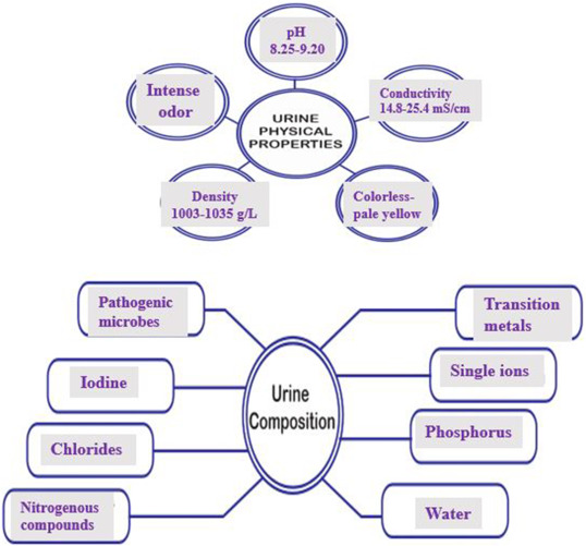
The composition also varies from country to country. For example, high income earning countries typically have the daily mean concentration of potassium as 2.2–2.7 g K/L, phosphorus as 0.74–2g P/L and nitrogen as 2.2–2.7 g K/L [5,7,8]. Among the components that are obtained from urine, 75–90% of nitrogen excreted are mainly in the form of urea with the remainder being released in form of uric acid, creatinine, and amino acid [7,8]. Urine also contains other ions like Na+, Cu2+, Mg2+, Cl− that are essentially required by plant for growth [9]. Apparently, human urine is preferably desired for use as fertilizer due to trace concentration of heavy metals in urines obtained from animal origin. Stored urine has different pH compared to the fresh urine. This is because under non-sterile environment, stored urine is hydrolyzed into bicarbonate and ammonia. Ammonia and carbon dioxide (from the bicarbonate) are released to the environment while the remaining bicarbonate raise the pH of the urine solution [10]. The physical properties and the composition of urine are summarized in Fig. 1.
 Fig. 1. Physical properties and composition of urine.
Some of the nitrogenous compounds found in urine are amino acids, ammonia, ammonium salts, urea, creatinine and uric acid [11]. Also, transition metals such as Cr, Mn, Fe, Co, Ni, Cu, Zn, Mo, Hg, Sb, Pb and As have been reportedly found in the urine [12]. Due to the presence of these numerous substances in the urine, it is important to comprehensively review how these substances can be captured and reused. Within the year 1864–2022, 543,512 articles were indexed on Scopus database when “urine” was used as the keywords (accessed on June 14, 2022). Some of these articles were used for writing this review. Other databases used are Google Scholar, Research Gate, PubMed Science, Springer Link, SciFinder and Science Direct.
2. Undesirable effects of urine
Compared to feces, urine is less dangerous but it also contains some disease-causing microbes. Examples of such microbes are L. interrogans, S. paratyphi, S. typhi and S. haematobium [13]. These microbes find their ways into living organisms to cause diseases. Deposition of urine in the soil alters the microbial activities and conditions of the soil, which consequently alters the way that nitrogen is being processed [14]. According to Singh et al. the urine of sheep disrupts the microbial communities of the soil [15]. Apart from the impacts on the microbial communities in the soil, it also affects the strength of the buildings. For instance, in developing nations where urine waste is not properly managed, urine leaches into the water used for construction works, which could diminish the strength of the concrete. The concrete that is cured with water containing high concentration of urine experiences reduced curing strength and this is as a result of lost in desirable properties when concretes are exposed to urine [16].
Urine in treated swimming water negatively affects the swimmer. This is because the uric acid present in the urine undergoes a reaction with chlorine in treated water to give trichloramine and cyanogen chloride, which are the same substances that are inhaled in swimming pools that results to itchy and redness of eyes [17]. Apart from this, the nitrogen released through urine could be processed into nitrogen gas, nitrous oxide, nitric oxide, nitrate or nitrite. These products also have significant impacts on the environment. For instance, N₂O is a greenhouse gas [18], while nitrate can cause eutrophication [19]. Urine-contamination has also been reported to affect the mobility of sperms [20]. For instance, Dreanno ab et al. [21], observed that the velocity of sperm reduced from 230 μm s−1 to 160 μm s−1 when it exists in a system containing 30% urine. Apart from the mobility, there was an observed decrease in the endogenous ATP that was stored in the sperm and fertilization capabilities when it existed in the environment of urine.
In addition to direct impacts of urine, the odor from urine has been reported to negatively impact on the behavior of livestock. It usually cause cattle to experience stretched locomotion and their attitude to breathing-in air [22]. The aversive odor from the urine of cat has been discovered to trigger avoidance character and initiate a freezing response in rats [23]. Finally, discoveries of SARS-CoV-2 RNA in the urine samples of patients that were infected with COVID-19 and from the wastewater of the community of the infected patients has heightened the possibility of transmission from urine and urine-polluted water [[24], [25], [26]]. This has raised a serious concern on the need to use both destructive and non-destructive conventional methods for the treatment of urine-contaminated water [[27], [28], [29]].
2.1. Detection of substances in urine
Urine is a good diagnostic tool for investigating the use and abuse of substances in some situations. This is important in forensic toxicology, workplace testing and treatment programs [30,31]. Several substances have been detected in urine by using different detection techniques. For instance, ten psychoactive products of plant (yohimbine, scopolamine, psilocin, lysergic acid amide, ibogaine, harmine, harmaline, ephedrine, N,N-dimethyltryptamine and atropine) have been detected in urine of human. Electrospray ionization detector was used for their sensing after reverse phase chromatographic techniques has been used for the separation of the substances from urine samples [32]. 4-methylmethcathinone, which is also clinically-active psychoactive substances, was detected in the urine of human through a paper-based colorimetric detector [33]. The psychometric substances that have been detected in the urine are numerous. Bijlsma et al. [34], reviewed various methods of identifying, detecting and quantifying novel psychoactive substances and their metabolites in the urine. Apart from these psychometric substances, detection and quantification of protein in human urine have been reviewed [35,36]. Other substances that have been detected from urine and their methods of detections are shown in Table 1.
Table 1. Detection of substances in urine.
| Source of urine | Substance(s) detected in urine | Detection/determination method | Separation method/material used | Detection limit | Ref. |
|---|---|---|---|---|---|
| Swine urine | Sulfathiazole and sulfadiazine antibiotics | Surface enhanced Raman spectroscopy | Gold nanoparticles combined with amine and carbon compounds | – | [37] |
| Human urine | Uric acid | Electrochemical sensor | – | 5 nM | [38] |
| Human urine | Per- and polyfluoroalkyl substances | Triple quadrupole mass spectrometer | Solid-phase extraction | – | [39] |
| Human urine | Uric acid | Enzyme-free fluorescent sensor | – | 0.6 μM | [40] |
| Human urine | Human serum albumin | BODIPY-based fluorescence probe | Column chromatography | 1.89 μg/mL | [41] |
| Human urine | Human serum albumin | Fluorimetric method | – | 0.1 μM | [42] |
| Human urine | Chlamydia trachomatis | Fluorescence detection | – | 40 μL | [43] |
| Human urine | Opisthorchis viverrini antigen | Fluorescence detection | – | 33 ng/mL | [44] |
| Human urine | Tizanidine hydrochloride | Square wave voltammetry | Adsorption | 1.15 × 10⁻⁸ M | [45] |
| Human urine | Adenosine | Fluorescence detection | – | 6.87 μM | [46] |
| Swine urine | β-agonists | Liquid chromatography-tandem mass spectrometry | Solvent extraction method | – | [47] |
| Human urine | Bismuth (III) ion | Fluorescence detection | – | 1 pM | [48] |
| Human urine | Hormones | Quadrupole time-of-flight | Chromatographic separation | 0.25–500 ng/mL | [49] |
| Pig urine | Phenothiazines | Chemiluminescence | – | 2–5 pg/mL | [50] |
| Human urine | Cadmium (II) ion | Voltammetric technique | – | 324 nM for male urine and 5.85 nM for female urine | [51] |
| Rat urine | Homogentisic acid | Electrochemical detection | Membrane filtration | 1.2 ng/mL | [52] |
3. Applications of urine
3.1. Application of urine in synthesis
Human urine and that from different animals have been recently utilized in the synthesis of nanomaterials. Urine of cow has been used to produce silver nanoparticles and the urine served as both capping and reducing agent. The presence of urea in urine made it a useful agents for the synthesis and was able to stabilize the silver nanoparticles produced [53]. The need to boost the amount of urea in the system led to the introduction of honey into urine for nanoparticle synthesis. The high percentage of urea in honey (6% unlike cow urine that has 2.6%) was the reason behind its usefulness in nanoparticles formation [54,55]. These mixture was used as capping agent in the synthesis of ZnO nanoparticles through combustion techniques. The honey also acted as a chelating agent in the synthetic system. When the particle size of ZnO obtained from pure cow urine and pure honey were compared under similar combustion conditions, there was marked difference in their particle size. The size of the nanoparticle produced was smaller when pure urine was used as the capping agent compared with the size obtained from pure honey [55]. Removal of oxygen from graphene oxide to form graphene nanosheet and simultaneous nitrogen doping of the formed graphene nanosheet has been achieved by using cow urine [56]. The process involved in using cow urine to achieve these reactions is summarized in Fig. 2. The method gave a high yield of nitrogen-doped graphene nanosheet.
 Fig. 2. Conversion of graphene oxide to graphene nanosheets and N-doping of graphene nanosheets by using cow urine. Reprinted with permission from Ref. [56]. Copyright (2017), Bentham Science.
Apart from using combustion method, cow urine has also been used as stabilizing and reducing agents through a biogenic method for the synthesis of ZnO nanoparticles [57]. It is also useful in the precipitation of calcite through a reaction involving hydrolysis of urea [58]. Other materials made from cow urine are shown in Table 2. Reduction of graphene oxide to graphene has also been achieved by using the cow urine as the reducing agent. The reduction was assisted with high temperature and it was observed that the efficiency of reduction increases with increasing temperature until it reaches a temperature of 140 °C [59]. This is a green synthetic route which minimized the quantity of harmful chemicals that would have been required for the reductive synthesis [60]. Graphene oxide grafted with zwitterionic polymer was used to enrich N-glycopeptides leading to the synthesis of a novel hydrophilic material. The source of the N-glycopeptides used for the synthesis was urine and it was also obtained from the urine of healthy human being and the lung adenocarcinoma patients. The N-glycopeptides that was extracted from the urine of patients with lung adenocarcinoma was compared with the one extracted from the urine of healthy person. The amount of the synthesized material obtained from the urine of patients suffering from lung adenocarcinoma was more than the one obtained from healthy person [61]. Although, the urine obtained from the two sources were useful for the synthesis.
Table 2. Synthesis of nanoparticles by using cow urine.
| Description of the cow used | Nanoparticles prepared | Chemical used | Particle size | Application of synthesized nanoparticles | Refs. |
|---|---|---|---|---|---|
| Bos taurus | Pd | PdCl2 and cetyl trimethyl ammonium bromide | – | Antioxidant and antimicrobial agents | [62] |
| Indian cow and Jersey cow | Ag | AgNO3 | 11–20 nm | Antibacterial agent against Klebsiella pneumonia, Salmonella abony, Escherichia coli, Staphylococcus aureus, Bacillus subtilisand Streptococcus epidermis, | [63] |
| Cow(species unspecified) | Cu | CuSO4 | 98 nm | Antioxidants and cytotoxicity evaluation | [64] |
| cows of the Tumkur city | Ag2O | AgNO3 | 11 nm | Antibacteria, photoluminiscence and photocatalytic applications | [65] |
| Cow(species unspecified) | CuFe2O4 | Cu(NO3)2·6H2O and Fe(NO3)2·9H2O | 13.7–18.2 nm | Magnetic material | [66] |
| Amrithmahal cattle | Ag | AgNO3 | 47.8 nm | Inactivation of Staphylococcus aureus and Escherichia coli | [53] |
Human urine has also been used for materials synthesis. Zhuang et al. [67], reported the use of human urine as a precursor in the preparation of graphitic carbon nitride nanodots. The synthesis was achieved at 200 °C within 2 h using an autoclave. Doped porous carbon (URC) has also been produced from urine [68]. The synthesis was possible after the elimination of halite and sylvite present in the urine through acid treatment followed by carbonization under nitrogen. The entire synthesis process has been summarized in Fig. 3. Apart from synthesis of materials from human urine, doping of nanomaterials has also been achieved by using human urine as the dopant. For instance, the phosphorus in the upcycling urine was used to replace titanium in a titanium oxide lattice. This significantly changed the band gap, surface chemistry and morphology of the titanium oxide compared with the undoped titanium oxide [69].
 Fig. 3. Schematic illustration of the synthesis of porous carbon (URC) from human urine. Step-1: The yellowish brown deposit was obtained from urine in the dried form. Step-2: carbonization of the dried deposit in an inert atmosphere. Step-3: washing and etching by using dilute acid to remove the mixture of rock that generates pores in the structure of carbon. Reprinted fromRef. [68]. Open Access published by Scientific Report.
The synthesis of organic compounds using human urines have also been reported. Siddiqui et al. [70], synthesized biscoumarin derivatives, benzypyrazolylcoumarin, 1,8-di-oxo-octahydroxanthenes and chromenes by making use of the carbon content of human urine (HUC). A one-pot multi-component process involving aqueous ethanol as the solvent media was used in the reaction and aromatic aldehyde (2) was a common reagent in the entire synthesis process. This urine-based synthesis is simple and gave good yield. The mechanism of the entire synthesis process has been detailed in Fig. 4.
 Fig. 4. Synthesis of chromene derivatives (4), xanthene derivatives (5), benzyl pyrazolyl coumarin derivatives (9) and bis-coumarin derivative (10) with the aid of the human urine carbon (HUC) in ethanolic solution. Reprinted from Ref. [70]. Open Access published by MDPI.
3.2. Hydrogen generation from urine
Hydrogen and its compounds are very important for the sustenance of the ecosystem. However, hydrogen atoms are short-lived because they combine fast with oxygen to form water molecules on which biotic components survive [71]. Hydrogen has an exorbitant energy content, and as such, has been considered as an alternative fuel source with energy content of about triple that of gasoline [72]. It is non-toxic, tasteless, odorless, and a light gas [72]. These properties qualify hydrogen as a good alternative fuel source. Other properties of hydrogen that make it a suitable fuel substitute are high auto-ignition temperature, minimum ignition energy in the air, and wide flammability limits with atmospheric air, among others [73]. With the rapid increase in energy need due to the increase in industrialization, hydrogen holds promise for the future energy resources in the world as a renewable energy source. Further, the production of hydrogen plays significant role in economic enhancement. Since the need for energy has become a challenge, investigation into the possible production of hydrogen from biowaste, such as urine [74] has continued to gain interest. Fortunately, humans discharge approximately 2500 mL of urine per day; thus, contributes to a huge volume of liquid waste [75,76].
A number of studies have reported some methods used in the generation of hydrogen as a source of energy. An on-demand hydrogen generation from wastewater and urine was reported by Elitzur et al., [77]. Activated aluminumpowder was employed in the generation of hydrogen from urine, which generated approximately 200–600 mL/min/g Al of H2 [77]. The production of hydrogen directly from urine through urea electrolysis in the presence of a nickel catalyst was an earlier reported method of synthesis [78]. However, more studies are required for this direct method.
Furthermore, zero-valent aluminum nanoparticles, which were previously used in wastewater treatment to remove organic pollutants and heavy metals, have been employed in the generation of hydrogen via hydrolysis [[79], [80], [81]]. However, the susceptibility of zero-valent aluminium nanoparticles to passivation via the formation of alumina layer on the surface causes a reduction in the reactivity and impedes its applications in the environment [79]. Malek et al. [79], recently developed a method involving the formation of aluminum nanoparticles via an in situ process in urine via the reduction of aluminum salt (Al2(SO4)3·16H2O) using sodium borohydride. The in situ method included the instantaneous reaction between the formed aluminum nanoparticles and urine to produce hydrogen. This method is summarized in a schematic diagram presented in Fig. 5. A previously reported method [79] bypassed the Al2O3-passivation layer-related challenge regarding hydrogen production and overcomes material storage and hydrogen transportation-related problems, since the reaction is in situ in nature.
 Fig. 5. Production of hydrogen via the reduction of aluminum salt in urine using NaBH4.
3.3. Treatment with urine (urotherapy)
Urine has been used in medical treatment, and Camel (Camelus dromedaries)urine is one of the animal urines that has been extensively used for therapeutic purposes [82]. Camel urine gained relevance in urotherapy because pharmacological assessment revealed that no toxic risk is associated with its usage for treatment [83]. Camel urine is a good anticlastogenic agent that has shown good application as potent target for the drugs [83]. The utilization of camel urine as anticancer, antiplatelet, hepatoprotective and gastroprotective agents has been reported [82,84]. Its anticancer properties could be linked to its toxicity against HeLa cells, osteosarcoma and leukemic cells of human. Inhibition of c-tumors in onion as a result of the presence of camel urine has been reported [85]. The urines of different animals have been used for the treatment of mental illness, leprosy, leucoderma, amenorrhea, haemorrhoids, poison, tuberculosis, flatulence, loss of appetite, abdominal tumor, anaemia, colic, abdominal enlargements and dropsy. Animals whose urine have been used for these treatments include donkey, camel, horse, elephant, buffalo, sheep and goat [86,87].
The presence of different constituents in cow urine makes it useful for several therapeutic purposes. For instance, Allantoin in cow urine makes it a good substrate in aiding wound healing [88]. The presence of copper and gold hydroxide in cow urine makes it function as antipoison, thus, it is used in the elimination of toxic effects of residues from medicines [89]. Also, its hippuric acid, (a typical component of urine that increases with increase in consumption of phenolic compounds such as tea, fruit juices and wine), phosphates and uric acid makes it useful as diuretic agent. The high nitrogen concentration makes it useful in stimulating the kidney. In fact, cow urine taken orally has been reported to boost immune system, improve memory, relieve tension and lower the level of cholesterol in the body [88,89]. Human urine has also been used for the treatment of allergic diseases and autoimmune, by the collection of the patient's urine and processing it to obtain the peptide fraction of the urine. This was then administered to the patient after conditioning [90].
3.4. Antimicrobial applications of urine
Camel urine is a good antibacterial agent. Its potency is higher compared to cattle urine, and this is due to its high alkalinity, high salt concentration and low uric acid concentration. This composition is linked to the feeding habits and the type of plants that camel consumes. Camels prefer to feed on grasses containing high minerals concentration which cattles usually avoid [91]. Antifungal potential of camel urine has also been reported [86]. Though, it was more effective at high concentration than in diluted form. Urine of goat has also been reported as antibacterial agents. For instance, the peptides extracted from goat urine displayed a marked inhibition zone against Escheria coli and Salmonela aureus. So, the cationic urinary peptides extracted from goat is a potent antimicrobial agent which can be used to control various bacterial infections [92]. Urine from cow has also been used as antibacterial agents against Staphylicoccus aerus, Pseudomonas aeruginosa, Bacillus cereus, Escherishia coli and Aeromonas hydrophila, which were extracted from fish and the cow urine was effective against all the tested bacteria strains [93]. Both goat and cow urine distillates have been reported as effective substances against certain dental pathogens (Lactobacillus acidophilus and Streptococcus mutans). The goat urine displayed least minimum inhibitory concentration values of 5 μg/ml against these microbes [94]. In fact, the urine of these two ruminant animals have been discovered to be effective against fungi (C. falcatum), which is responsible for red rot disease in sugar cane [95]. Relatedly, the urine of animals such as buffalo, goat and dairy cow have been discovered to have antioxidant activities [96].
3.5. Urine as source of useful stem cells
3.5.1. Origin of urine-derived stem cells (USCs)
Knowledge of the origin of USCs will unravel the biological value of the multipotent cells in the urinary tract. Renal tissue derived stem cells have been sourced from the papilla and tubules. Moreover, glomerular epithelial cells could function as stem cells and differentiate into podocytes and call in the proximal convoluted tubule [[97], [98], [99], [100], [101]]. Some epithelial cells could lose their polarity and transform into mesenchymal cells. The resulting multipotent stromal cells could differentiate into various types of cells that could be used in the repair and regeneration of the renal tissue. Several evidences indicate that USCs possibly originate from the epithelial cells of the glomerular parietal. Studies have shown that USCs in urine samples in the superior portion of the urinary tract are similar in terms of structure, pattern of growth, differentiation ability, and cell phenotypes to voided USCs. This suggests that USCs in urine sample originate from the upper portion of the urinary system [102,103].
3.5.2. Characteristics of urine-derived cells
USCs express different markers for pluripotent stem cell (PSCs), pericyte and mesenchymal stem cells (MSCs). Urine-derived stem cells have been noted to express some prericyte markers, among which are CD-224 and CD-146. Specific MSC markers such as CD-44, -73, −90, and −105 are known to be expressed by USC [104]. Kruppel-like and octamer-binding transcription factors are notable among the markers USCs express for PSCs [102]. Moreover, USCs express epithelial cell adhesion and neural cell adhesion molecules, and sine oculis homeobox homologue 2, which suggest that these cells originated from the renal tissue [105,106]. In contrast to other widely used stem cells isolated from umbilical cord, bone marrow, and adipose tissues [107,108], USCs have high expandability. Asides, USCs with a mean doubling time of about a day, may attain about 70 population doublings. However, stem cells that are urine-based are known to have a doubling time of more than 24 h. Asides, complexity and considerable time have been associated with their isolation, culture, and sample processing.
Like induced pluripotent and embryonic stem cells, USCs are multipotent [109]. They have the ability to generate cells that originate from the three germ layers. Moreover, USCs secretes several growth factors of vascular, hepatic, fibroblast and platelet origins [110]. These growth factors with angiogenic and immunoregulatory function have been implicated in the vascularisation of USCs-derived cells, which on successful transplantation could boost the host immune system. It should be added that USCs could be used in tissue reparative procedure and in the treatment of several degenerative diseases.
3.5.3. Therapeutic uses of urine-derived stem cells
3.5.3.1. Liver reconstruction
Liver failure has affected a large percentage of people in different countries of the world. Organ transplantation is considered as a viable option for the management of this condition, especially, the acute case [111], Most patients lost their lives as a result of insufficiency of donor tissue or immune reaction due to the transplanted hepatic cells. Therefore, the provision of patient-specific hepatic cells will be a possible alternative for transplantation. Small molecules have been used to transdifferentiate renal cells into insulin-secreting cells [112]. The efficiency of transdifferentiation is guaranteed if the precursor cells originate from the same germ layer as the transdifferentiated cell [111]. The kidney cells highly expresses Sox-17, an endoderm marker, therefore, the transdifferentiation of urine-derived cells to endoderm-derived cells will be both easy and efficient. This is because the transdifferentiated and renal cells will share the same pedigree. Hence, isolated cells in the urine can be transdifferentiated into hepatic cells. These cells behave like mesenchymal cells and are known for their potency in generating hepatic cells in vitro [113,114].
3.5.3.2. Diabetes treatment
The incidence rate of diabetes world-wide is higher than other chronic disease conditions [115]. Over the years, tissues obtained from cadavers have been used in islet cells transplantation. However, this has been attended with insufficiency of cells required. Moreover, the recipients require a life-long immunosuppressant so that the grafted tissue will survive [116]. At the moment, larger numbers of β-cells are required for a successful transplantation procedure [117]. With the use of USCs these numbers could be easily achieved. In addition, small molecules have been used to differentiate renal cells into insulin-secreting cells [112]. An in-vivo study reveals that USCs isolated from healthy individuals could be differentiated into pancreas homeobox 1 cells [118] and so helps to abate the symptoms of diabetes. It is desirable that a similar study would be performed with USCs isolated from diabetic patients. This will clarify if there are significant differences in the cells isolated from diabetic subjects in terms of morphology, efficiency of isolation, and gene expression of markers. Such knowledge will guess our quest in the clinical application of this procedure. The ease of collection of USCs, coupled with minimal effort to culture these cells, and their efficient transformation into pancreatic beta cells could revolutionized changes in the management of diabetes.
3.5.3.3. Cardiac repair
Improper functioning of the cardiac tissue as a result of damages to the heart muscles eventually results in heart failure [119] Induced pluripotent stem cells (iPSC) and embryonic stem cells (ESCs) could differentiate under suitable condition to cardiomyocytes [120] However, these stem cells are difficult to obtain; moreover, the full-differentiation of the cells requires a lot of time. In contrast, USCs are easily isolated and could be differentiated without much effort in vitro [121]. Differentiated muscle cells from USCs have demonstrated an enhanced expression of surface markers and myofibrillar proteins, such as myogenin, myogenic factor 5, and myogenic differentiation factor [121]. Further investigations should be carried out on how to optimize the procedure to generate functional cardiac muscle cells from USCs. Expectations are high that with efficient screening of molecules, USCs could be differentiated into well-functional cardiomyocytes, which could be used to repair the heart defects.
3.5.3.4. Neuroregeneration
There are limited clinical trials on the use of nerve cells derived from stem cell for the treatment of disorders in the brain tissue. In-vitro studies have revealed the production of potent nerve cells from differentiated PSCs or neural stem cells [122]. However, it should be noted that PSCs-based protocols take about 8 weeks to generate functional nerve cells [123]. Moreover, post-transplantation in vivo, these cells lose their biological function as a result of damaged dendrites after being separated from culture media. However, this difficulty could be prevented by transplantating immature cells that could undergo maturation process in a suitable media. Studies have shown that USCs cultured on laminin-treated plates, with growth factors treatments efficiently convert into nerve cells [124]. Immature cells cultured in biomaterials can be transplanted in vivo with insignificant loss in their structural component, resulting in easy integration and maturation process. Cheng and fellows generated neural progenitor cells from renal cells. These cells were noted to be functionally potent both in vitro and in vivo [125]. Therefore, functional nerve cells can be produced in larger quantities and hence transplanted for neuroregenerative therapy, without any significant cell loss during the process.
3.6. Urine as a source of biomarkers in disease conditions
3.6.1. Urine as a source of biomarker for stroke
Stroke is neurodegenerative disease with attendant high morbidity and mortality as a result of sudden obstruction or bleeding of brain vasculature [126]. Presently, the condition is diagnosed with imaging techniques and observed based on clinical signs. The accuracy of computed tomography is about 82% after 6 h of impaired blood flow to the cerebral tissues. Contrarily, the false negative rate of magnetic resonance imaging could be significantly high; moreover, the technique cannot be used for subjects with implants [127].
Urinary proteomic markers for stroke were demonstrated for the first time with the aid of Two-dimensional gel electrophoresis (2-DE), which was used to assess the effects of salt administration on the protein molecules in urine and serum samples. In the study, several proteins, for examples transferrin, hemopexin, albumin, transthyretin, among others, were detected in the urine of the stroke-prone rats, prior to the manifestation of clinical signs of stroke after salt administration [128]. Furthermore, urinary metabolomics could be instrumental in the search for stroke bioindicators. Jung and colleagues noted that urine metabolome in healthy individuals is different from those observed in stroke patients [129]. Notable alterations in the metabolites found in the urine of stroke survivors include reduced concentrations of some biomoleculessuch as hippurate, glycine and dimethylamine. Reductions in these metabolic markers have been linked with deficiency in folic acid and excessive presence of homocysteine, which have been linked with an elevated risk of stroke and cognitive inadequacies [130].
3.6.2. Urine as a source of biomarker for Alzheimer's disease (AD)
AD is a degenerative brain disorder, with high incidence rate in several countries [131]. Clinical confirmation of the condition is dependent on neuropsychological evaluation alongside with determination of bioindicators in the cerebrospinal fluid [132]. Early diagnosis should rather be aimed at prior to the onset of irreversible damage of the brain tissues and decline in mental state. Hence, it is pertinent that an accurate and efficient method for early diagnosis of AD is sought. Fukuhara and colleagues used NMR to analyze the metabolome in the urine sample of Tg2576 transgenic AD mice [133], and they observed that biomolecules such as tyrosine, homogentisate, and 3-Hydroxykynurenine are effective biomarkers of AD before the onset of dementia. However, they added that late-stage AD was characterized by other biomarkers e.g. 2-oxoglutarate, citrate, urea, trimethylamine, 1-methylnicotinamide, trigonelline, among others. In another study, urinary metabolomic biomarkers were used in an experiment in which transgenic mice model of AD was compared to healthy mice [134]. The level of urine metabolites was observed to be significantly different between the normal and the transgenic AD mice at 15–17 weeks after birth. The noted change became more noticeable after a period of about six months and was accompanied with observable alterations in the AD mice model. The researchers identified desaminotyrosine, taurine, methionine, among others as promising biomarkers in the urine for the diagnosis of AD.
3.6.3. Urine as a source of biomarker for breast cancer
Bioindicators for prostate cancer can be assessed in urine samples. This is because it is a urogenital disease. However, breast cancer, which is a non-urogenital can also be diagnosed by biochemical analysis of urine sample. Breast cancer is the commonest cancer among women. Yearly, about 1.4 million new cases and about 500,000 mortality rate have been reported among women worldwide [135]. Nevertheless, early detection of breast cancer could significantly increase the survival rate of subjects [136]. Specifically, clinical breast examination and mammography have been recommended for early diagnosis of this condition. Studies have shown that mammography reduces mortality as a result of breast cancer by 15%–20% [137]. This shows that some cases cancer of the breast cannot be detected by the technique. The National Academy of Clinical Biochemistry have recommended some biomarkers for the evaluation of breast cancer [138]. Among these include progesterone and estrogen receptors, urokinase plasminogen activator, cancer antigen 15–3, plasminogen activator inhibitor 1, to mention a few. These markers have been noted to be of use in predicting response to treatment; however, they proved to have little potential for timely detection of cancer [139]. Previous studies have reported biomarkers in the urine for detection of breast cancer [140]. Notable among these are matrix metalloproteinases and ADAMS, which have been implicated in the process of tumor progression [141]. A study in which urine sample was used revealed that females with elevated levels of ADAM-12 and matrix metalloproteinase-9 are susceptible to precursors of cancer, such as hyperplasia and lobular carcinoma [142]. Reports showed that ADAM-12 expressed by cancer cells, hasten the progression of breast cancer by causing stromal cells apoptosis [143] and by destroying components of the extracellular matrix [144].
3.6.4. Urine as a source of biomarker for diabetes
Diabetes is a metabolic disease condition mainly characterized by hyperglycemia. If left unmanaged diabetes could result in complications diabetic nephropathy, neuropathy and retinopathy. The two types of diabetes have different pathogenesis. Type 1 and II diabetes mellitus (T1DM & T2DM) are known to be instigated by the autoimmune destruction of pancreatic beta cells and insulin receptor defects, respectively. Clinically, both types of diabetes are diagnosed by determination of haemoglobin A1C and fasting blood glucoselevels. Moreover, glucose tolerance test [145] is also being assessed. The International Diabetes Federation [146] reported that T2DM frequently goes undetected, resulting in further complications. Several occurrences of diabetes both in the developed and under-developed countries are not diagnosed, while those that are detected are often diagnosed lately, usually 4–7 years after the disease onset [147]. Therefore, efforts are channeled towards finding early onset biomarkers of the disease [148]. The urinary proteome of over 300 individuals were evaluated using Capillary electrophoresis–mass spectrometry. About 261 bioindicators were noted, which could be used to differentiate those with/without diabetes. The results in the aforementioned study were confirmed in a larger study [149] that included both diabetic subjects (type 1 and 2), and yet with comparable sensitivity and specificity of the results obtained. In these studies, the most acclaimed biomarkers of diabetes that could be detected in the urine include fragments of type I and type III collagen α-1, uromodulin, α1-antitrypsin, and fibrinogen. A significant decrease in collagen fragments was noted in the diabetic subjects, compared to the healthy controls. This reduction was more significant in T2DM patients, relative to those with T1DM, pointing to variations in the extracellular matrix remodeling.
3.7. Nutrient recovery from urine
The recovery of nutrients basically involves measures/technologies that are adopted for the isolation of nutrients in urine either by volume reduction or transformation into solids [150]. Such measures include, ammonia stripping, evaporation, selective adsorption of nutrients, struvite precipitation. A compendium of various approaches used for nutrient recovery is shown in Table 3.
Table 3. Summary of methods used for nutrient recovery in human urine with their merits and demerits.
| Nutrient recovered | Treatment measures | Efficiency (%) | Advantages | Limitations | References |
|---|---|---|---|---|---|
| Nitrogen, Phosphorus | Chemical | 91 | -Highly efficient and selective for substance recovery. |
|
[151] |
| precipitation | 85 | ||||
| Ammonium, Nitrate | Distillation and | 95 |
|
|
[152] |
| nitrification | |||||
| Phosphate | Chemical process ion exchange | 90–93 |
|
|
[153] |
| Phosphorus | Struvite precipitation | 94 |
|
|
[154] |
| Phosphate, Ammonium | Membrane separation | >90 |
-Need simple operational techniques.
|
|
[155] |
| 50–80 | |||||
| Phosphate | Urea hydrolysis | 82 |
|
|
[156] |
| Ammonium | Freezing | 90 |
|
|
[157] |
|
|||||
| Ammonia | Air stripping | 93 |
|
|
[158] |
| Ammonia | Bio-electro chemical systems. | 60 | Energy cos is low through the elimination of aeration. |
|
[159] |
3.7.1. Application of selective adsorption for nutrient recovery from urine
Selective adsorption method makes use of an adsorbent to extract nitrogen suitable for usage as a soil conditioner. It has been implemented to majorly recover nitrogen from urine by using a selective sorbent known as zeolite. Zeolite sorbent primarily comprises of alumina and silica and are both commercially/domestically available worldwide [160]. Significant recovery of phosphorus nutrients can also be achieved if used together with struvite precipitation. Highest recovery rates of nitrogen and phosphorus were obtained by dosage addition of MgO at 0.5 mg/L and zeolite dosage of 15 g/L with the supernatant concentration of nitrogen and phosphorus to be 1000 gNm−3 and 10 gpm−3 respectively [7,10,161]. Recovery of nitrogen is a selective adsorption process that actually depends on the ion exchange of the sorbent/resin [150]. The sorbent is fortified with electrically charged active groups, which are capable of adsorbing ions. The sorption of nitrogen to zeolite is dependent on its affinity for the active groups on the adsorbent. Upon adsorption of nitrogen, the sorbent were been used for conditioning the soil since they are useful material in improving both nutrient and water retention in soils having low nutrients [162,163]. Maurer et al. speculated that nitrogen (∼64–80%) in urine can be recovered via zeolite adsorption [7]. Other class of natural zeolites such as clinoptilolite has been selectively used for the adsorption of ammonium [7,161].
3.7.2. Struvite precipitation for nutrient recovery from urine
Struvite precipitation is a method for frequently recovering nitrogen and phosphorus-based nutrients by creating fertilizers as product in solid form [164]. It has been commonly reported for use in phosphorus recovery [7,159,165]. This technique is implemented in some wastewater treatment plants (WWTP) and pilot facilities [166]. Struvite is basically an orthophosphate with cations (magnesium and ammonium ions) and anion (phosphate) in equimolar ratios [165]. MgNH4PO4•6H2O is predominantly used in wastewater to convert two dominant nutrients (nitrogen and phosphorus) in solid form.
Increased struvite precipitation has been achieved by the addition of magnesium to urine. Upon addition, struvite precipitate can be filtered and washed with water followed by drying [160,167,168]. Struvite is preferentially selected due to its slower rate of nutrients release, less impurities compared to commercially used phosphate fertilizers and interestingly due to the sorption of essential elements (P, N and Mg2+) on the sorbent simultaneously [7,169]. Unlike in selective adsorption process, precipitation of struvite favors the recovery of phosphorus but only allows limited fraction of nitrogen to be formed. Ahmed et al. posited that the ratio of nitrogen to phosphorus in fresh urine is about 0.5 in struvite [159,170]. According to Ahmed et al. pure and dry struvite typically contains 5.7% nitrogen and 12.5% phosphorus [170]. Factors such as precipitation and drying processes are determinant factor on phosphorus concentration on struvite sorbent. Phosphorus in struvite sorbent was reported to be 100 times more concentrated than its composition in urine [159,171]. The general reaction of struvite precipitation follows the equation below with a molar ratio of 1:1:1.[165]NH4+ + HnPO43−n + 6H2O + Mg2+ → nH+ +MgNH4PO4·6H2O ↓ (n = 0, 1, or 2)
3.7.3. Application of evaporation method for nutrient recovery from urine
Evaporation is used for concentrating the nutrients in urine. It is commonly known as a volume reduction method. Since nitrogen and ammonia are major component of urine, nitrogen is stabilized prior to evaporation to prevent gaseous ammonia emissions and losses of nitrogen [159]. Water was evaporated by adjusting parameters such as temperature, hydrometry and pressure [172]. It can also be a simple volume reduction process with the final product in liquid form or may proceed until dehydration with the final product been solid. Evaporation is regarded as a straight-forward technology aimed at removing water of high quality from urine [7]. Effort has been applied to recover pure water from urine for space application. National Aeronautics and Space Administration (NASA) in 2005 planned to install urine processing plant in the international space station via the application of vapor compression distillation (VCD). More than 96% of water content with energy requirement of 277–396Mjm−3 for small-scale unit will be recovered [173]. In another investigation, Holland et al. applied lyophilization to obtain ice at −90 °C from frozen urine under vacuum [174]. Pertinent challenges associated with urine evaporation include loss of ammonia and amount of energy consumed. The loss of ammonia can be addressed by the use of non-hydrolyzed urine or acidification process while energy loss can be minimized by energy recovery. Most large scale pilot plants adopt VCD methods for this purpose [175]. Examples of other pilot scale designs incorporating both distillation and nitrification process are shown in Fig. 6 (A and B).